
Background
 |
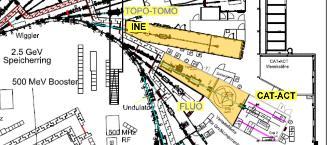
The INE Beamlines (INE-Beamline and the ACT station at the CAT-ACT Beamline) at the KIT-synchrotron are dedicated to actinide research with X-ray spectroscopic techniques. Both are operated by the Institute for Nuclear Waste Disposal (INE).
|
|
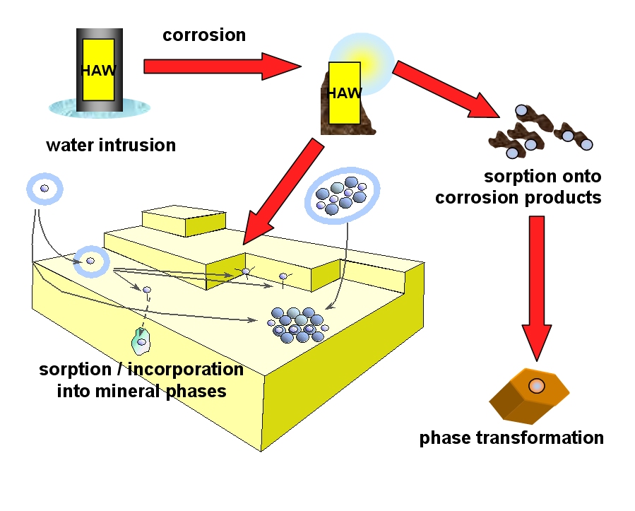
Schematic sketch visualizing some of the actinide mobilization / retention processes possibly following actinide release from corroded HAW. |
Research and development at INE are largely aimed at long term safety assessment of proposed deep geological repositories for high-level, heat producing nuclear waste (HAW) disposal. To ensure sound safety assessment, a molecular understanding of processes determinant in the fate of radionuclides, notably the actinides, and thermodynamic quantification is essential. Of central importance in such investigations is determination of actinide speciation (i.e., their chemical and physical form). X-ray spectroscopic methods have proved to be valuable tools for actinide speciation research. Investigations on non-fissile radioisotopes up to 106 times the legal exemption limit and fissile radioisotopes (Pu-239, U-235) up to 200mg, contained within two layers of protection, are possible. The synchrotron-based activities at the INE-Beamline are embedded in INE’s in-house research, thereby allowing a combination of analytical and instrumental methods, notably laser techniques and microscopic methods. |
In situ X-ray spectroscopic investigations on radioactive samples are routinely performed at the INE Beamlines for studying, e.g., actinide containing solid-water interface chemical reactions, not possible at other facilities. A special protocol for working with radioactive samples at both INE beamlines exists and is supervised by INE’s own radiation protection officers. The unique aspect of the INE Beamlines is its close proximity to INE’s active laboratories on-site KIT north campus. The design is for a multi-purpose beamline, where a number of methods are possible on one and the same sample.
The INE Beamlines serves three general functional areas:
- Research = user facility (INE in-house, HGF NUSAFE program, via strategic research cooperation with KIT-INE)
- Education and dissemination (maintaining nuclear competence)
- Development & upgrades (adaption of methods, serving user needs)
Layout
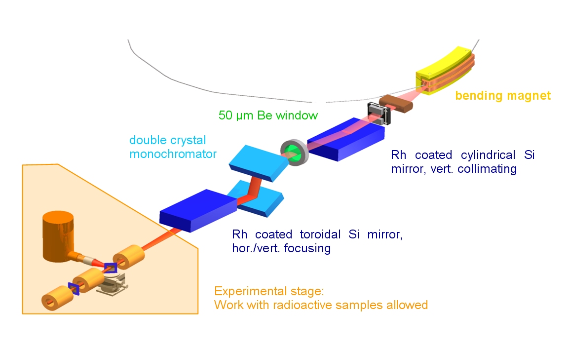
INE-Beamline – layout of optics and experimental stage.
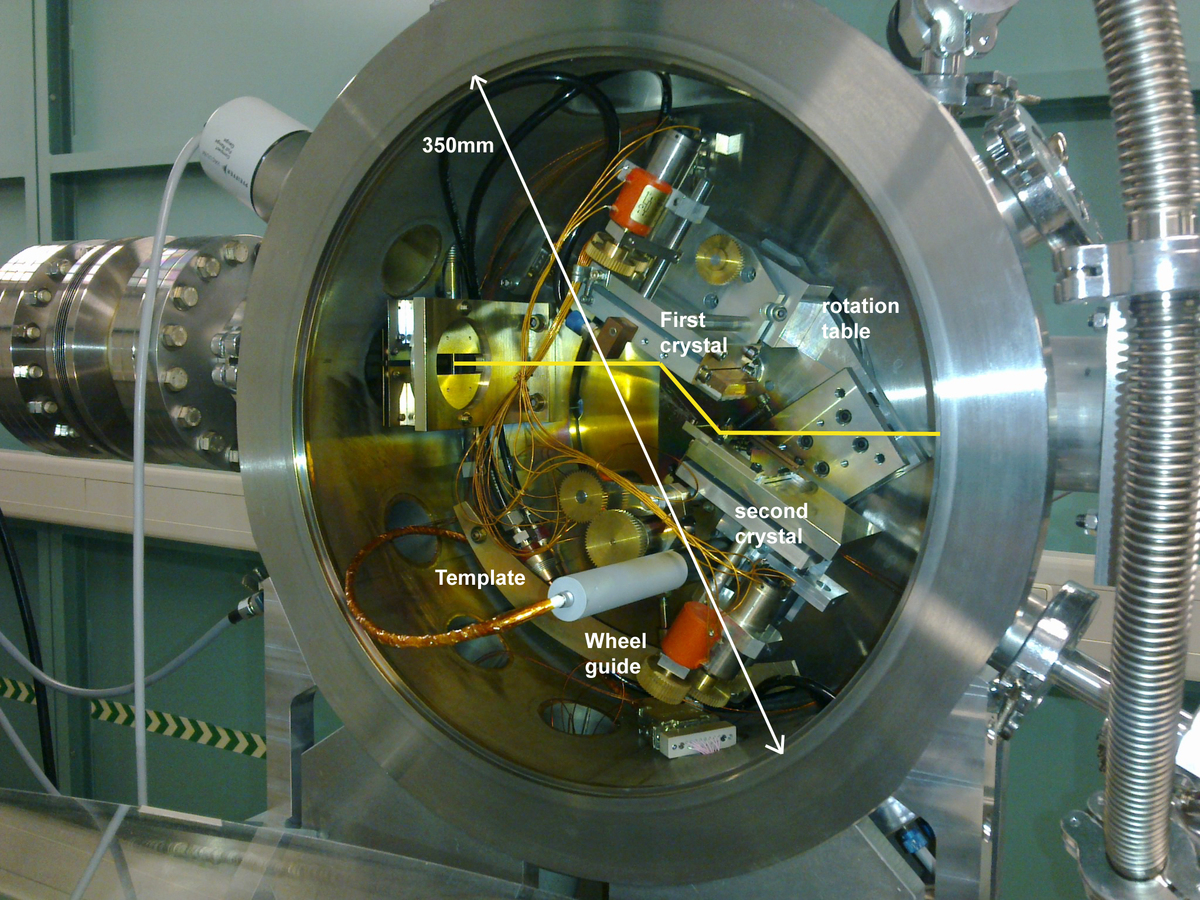
The compact design and pressure inside the DCM vacuum housing of ~10-6 mbar allows fast crystal changes without long pumping times (see figure beside).
The experimental station is flexibel designed to adapt different configuration setups as low energy with special chambers for sample, microfocus setup, etc... (see "experiment" for more information).
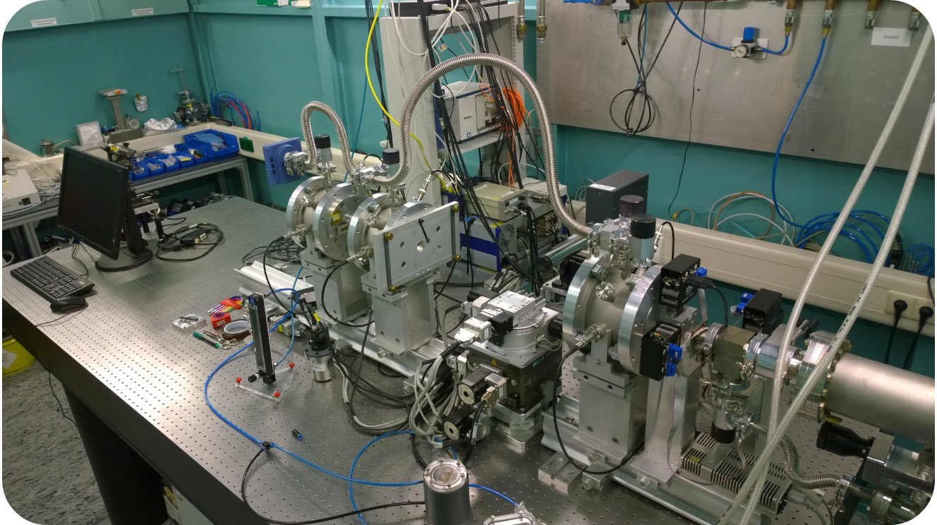
INE-Beamline standard setup
Experiment
Detectors
| Detector | Type | Main specs |
| Fluorescence |
4 element SDD Vortex (Hitachi, USA)
Vortex-60EX SDD (Hitachi, USA) |
130 eV at 6 keV; 45 mm2 active area/element; 25 µm Be window
130 eV at 6 keV; 50 mm2 active area; 25 µm Be window |
| Absorption |
3 ionization chambers (Poikat / Hamburg low-energy set-up with single entrance window (Oken Ltd. ionization chambers, Japan) |
window 12.5 or 25 µm KAPTON, FEMTO low current amplifiers DLPCA-200 ionization chambers and sample chamber form windowless volume – gas tight insertion of SDD into sample chamber for high quality fluorescence spectra |
| Area detector |
sCMOS (Photonics Science , England) |
window 156mm diameter active area 61.44 X 61.44 mm2 - pixel size 10µm x 10 µm resolution 6144 x 6144 (37,7 Megapixels) 2 removable scintillators available : GdOS 40µm thickness and CsI:Tl 100µm thickness |
Sample holder (HUBER goniometer):
| Movement | Range |
| z stage | 40 mm; 40 nm/step |
| phi circle | 360°; 0.0001°/step |
| psi cradle | 30°; 0.0001°/step |
| theta cradle | 30°; 0.0001°/step |
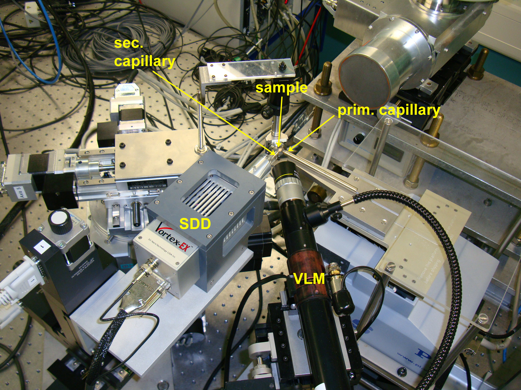
Setup for confocal measurements with a µ-focused beam at the INE-Beamline.
XYZ Sample positioner (IBG-2 design):
Alternative sample positioner with larger travel ranges designed by KIT-IBG-2 using Phytron controllers.
| Movement | Range |
| z stage | 150 mm; |
| y stage | +/- 72mm; 20µm /step |
| x stage (along beam) | +/- 22mm; 20µm /step |
Sample environment
• Standard holder for transmission and fluorescence measurements of radioactive and non-radioactive samples, e.g., 400 µl and 2 ml PE-tubes, pellets.
• Special sample chambers for grazing incidence measurements of flat samples (up to 3 inch diameter)
• Liquid sample cell for the investigation of redox labile systems under electrochemically controlled conditions
• High T / pressure cell setup
• Environment: atmosphere, inert gas (existing supplies: He, N2, Ar)
• Liquid N2 cryostat (OI OptistatDN) for low temperature measurements
• setup for tender X-ray measurements* (currently applicable down to the phosphorus K-edge at ∼2.14 keV)
* see Dardenne, K. et al. Inorg. Chem. 2021, 60, 12285−12298
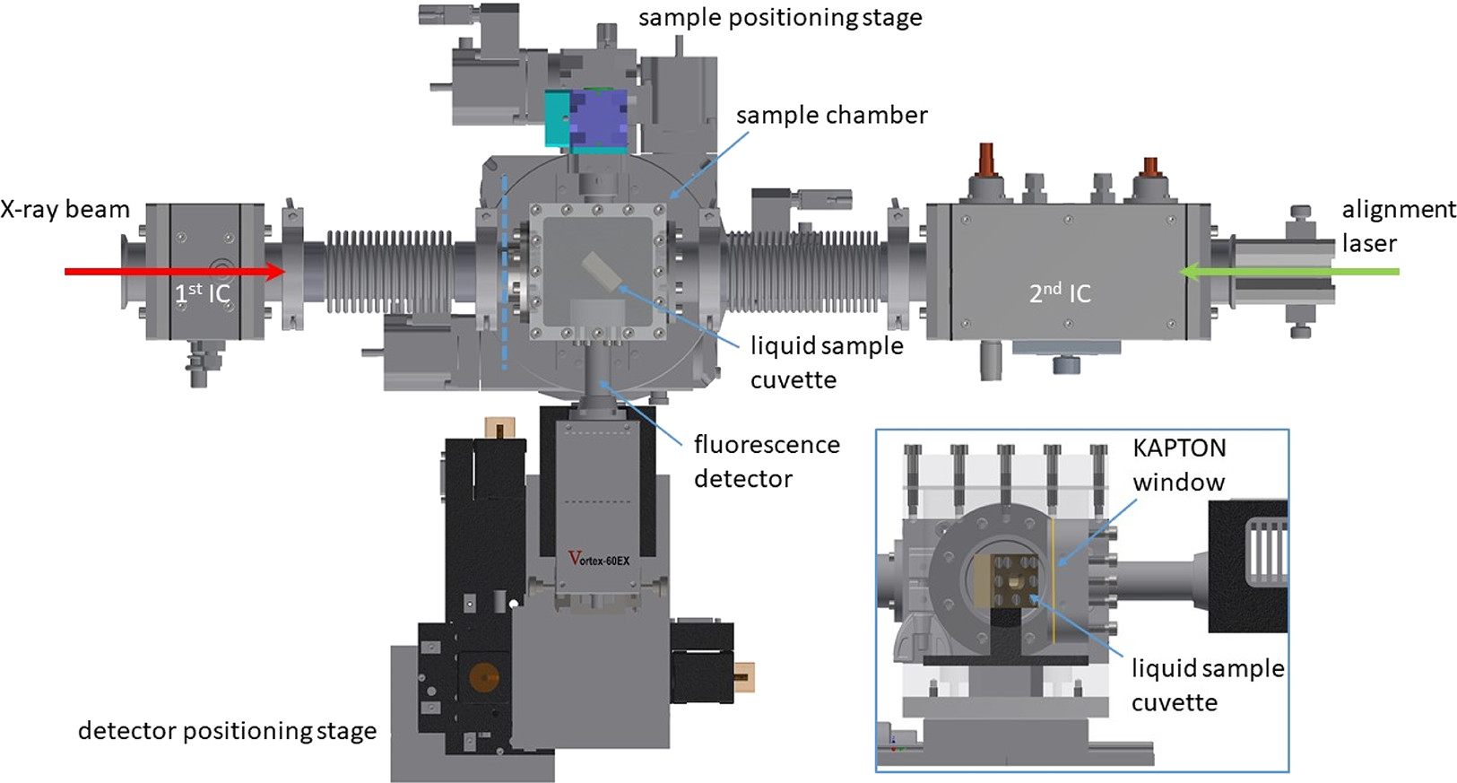
Tender X-ray setup (Inorg. Chem. 2021, 60, 16, 12285-12298).
Methods
| XAFS | characterization of bulk species by XANES/EXAFS |
| XAFS/XRD | characterization of phase - pair distribution relationships |
| XRF | measure elemental concentrations |
| Surface sensitive | with grazing incidence (GI) techniques |
| GI-XAFS | characterization of surface sorbed species |
| X-ray reflectivity | determination of surface layer thickness and roughness |
| Spatial resolution | with focused beam for ‘micro’ or µ-techniques |
| µ-XAFS | chemical state imaging |
| µ-XRF | elemental mapping |
| µ-XRD | identification and distribution mapping of phases |
| Combination of methods | combined X-ray methods or X-ray method combined with other techniques, e.g., laser, Raman or UV/VIS spectroscopy |
Characteristics
| Radioactive work | Infrastructure and licensing for activities 106 times the legal exemption limit as well as Pu-239 and U-235 up to 200mg. |
| Energy range | 2.1 keV - 25 keV (P to Pd K-edge, actinides up to Cf L3 edge) |
| Source | 1.5 T Bending magnet (Ec=6keV) |
| Optics | • Double crystal monochromator, water-cooled first crystal, mechanically coupled movement of the second crystal to ensure fixed exit, D-MOSTAB, exchangeable crystal pairs InSb(111), Si(111), Si(311), Ge(422) • Rh coated silicon mirrors (1st mirror vertical collimating, 2nd mirror vert. and horiz. focusing) • SESO X-ray beam position monitor • Optical microscope for sample positioning • Set of polycapillary half lenses (IfG) for confocal detection of sample fluorescence yield at ~25 µm spatial (3D) resolution • Single bounce capillary lens (IfG) with long focal distance for diffraction measurements at ~35 µm lateral resolution |
| Beam size at sample position |
~500µm × 500µm (standard beam size) ~25µm with secondary focusing optic |
| Flux at sample position | ~2×10¹⁰ photons/s at Zr K-/Pu L3-edges using Ge(422) |
| Experimental set-up/ sample positioning |
• Standard sample holders for radioactive samples, other dimensions can be accommodated |
| Experimental set-up/ detectors | • Ionization chambers for nominally high energies (transmission mode) • Ionization chambers for nominally low energies (Oken Ltd, Japan) • Setup for total electron yield (TEY) measurements • XRD-setup using area detector (sCMOS 37.7MP, Photonic science) • PIN diode to measure photon flux at sample position • Silicon drift detector 4 elements (Vortex, SII NanoTechnology) • Silicon drift detector (Vortex-60EX, SII NanoTechnology) • Fully digital fluorescence detector read-out (XIA) |
| Name | Tel. | |
|---|---|---|
| Dardenne, Kathy | +49 721 608-26669 | dardenne ∂does-not-exist.kit edu |
| Prüßmann, Tim | tim pruessmann ∂does-not-exist.kit edu | |
| 1 additional person visible within KIT only. | ||
| Name | Tel. | |
|---|---|---|
| Dardenne, Kathy | +49 721 608-26669 | dardenne ∂does-not-exist.kit edu |
| Prüßmann, Tim | tim pruessmann ∂does-not-exist.kit edu | |
| 2 additional persons visible within KIT only. | ||