Surface Analysis and Microscopy
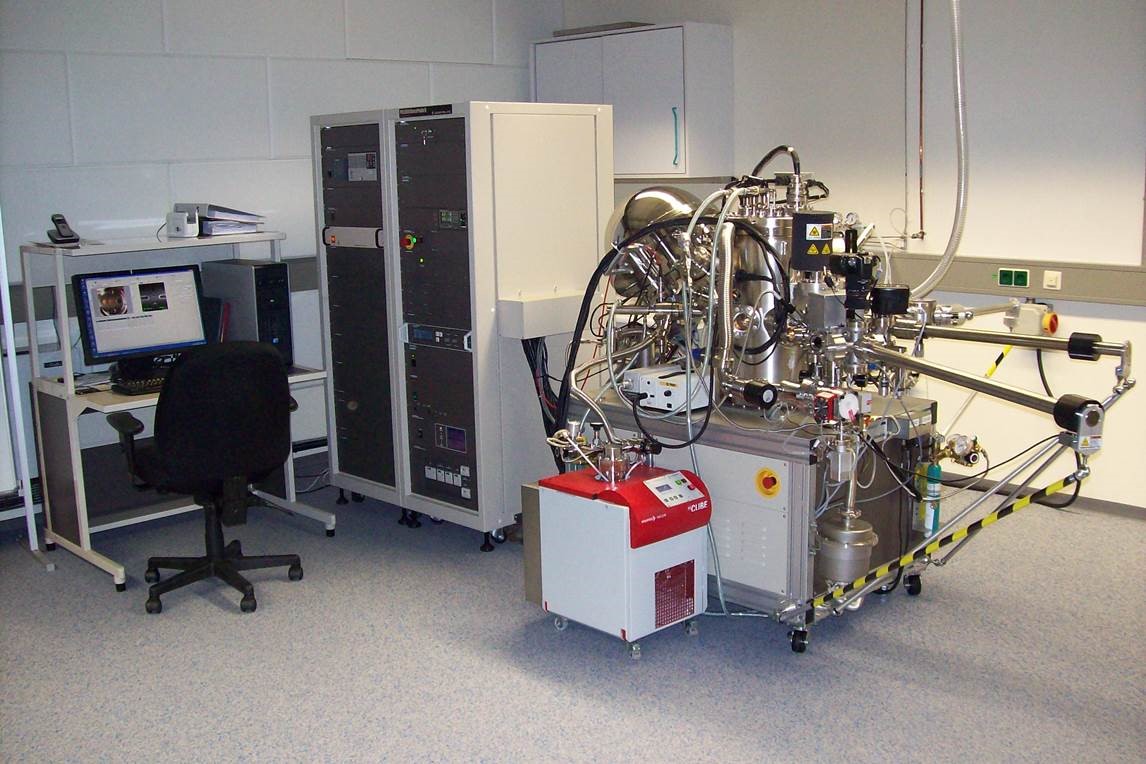
Chemical interaction of a solid material with its environment takes place at its surface, which mostly results in the formation of an interface phase or a layer with a chemical composition differing from that of the bulk. The thickness of such layers may range from monolayers to some nanometers. Knowledge of surface layer properties is essential for understanding of chemical processes occurring at the surface and relevant for the safety case study of a nuclear waste repository. X-ray photoelectron spectroscopy (XPS) gives insight into elemental composition, valence, and chemical bonding states of the outermost atomic layers of a sample. Examples are the interaction of dissolved radionuclides with surfaces of corrosion products, colloids of the groundwater, and mineral phases by redox reactions, sorption or incorporation.
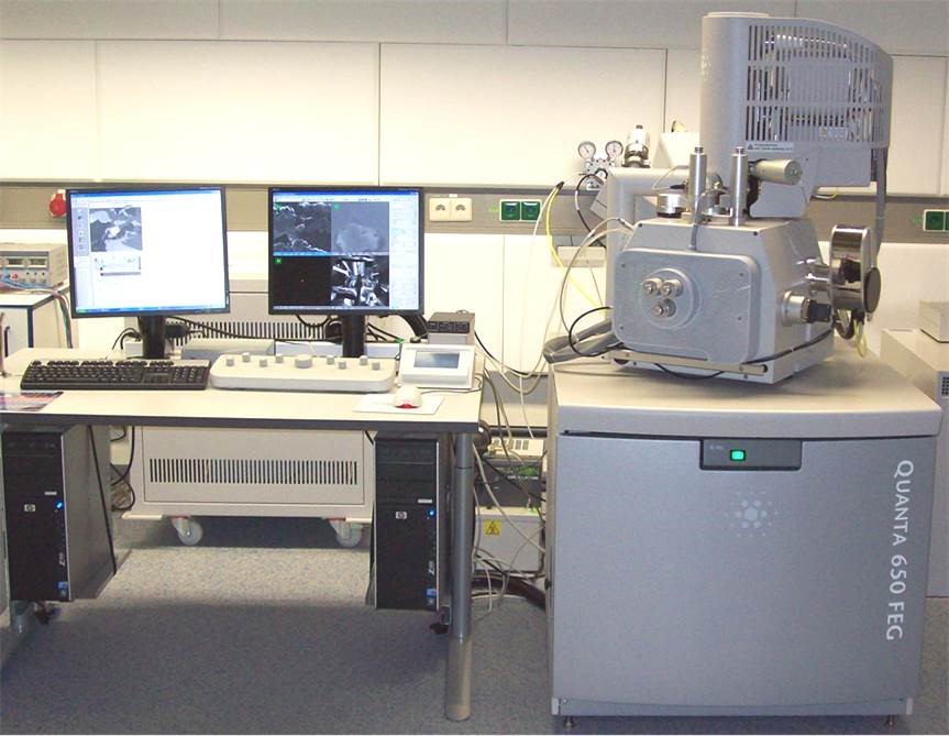
Analyses of solid surfaces at high lateral resolution is performed by environmental scanning electron microscopy (ESEM). In SEM, a fine electron probe is focused on the surface of a sample and scanned in a two-dimensional raster. At each point, various emitted signals can be detected and contribute to a corresponding point in an image. So-called secondary electrons (SE) enable the observation of surface topography with a lateral resolution down to few nanometers, whereas images produced by detection of elastically backscattered electrons (BSE) present a material contrast. Furthermore, the detection of characteristic X-rays allows determination of atomic concentrations at a micrometer scale (energy-dispersive x-ray spectroscopy, EDX).
Mineral phases and actinide compounds are also analyzed by Raman spectromicroscopy (Bruker Senterrra). Solids can be analyzed in-situ in aqueous solutions.
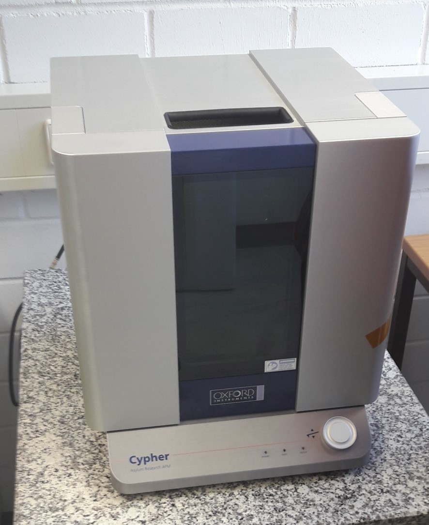
Atomic force microscopy (AFM) is used to map surface structures and chemical reactions at mineral-water interfaces like e.g. mineral dissolution or -precipitation in-situ with high spatial resolution (< 100 pm vertical and ca. 10 nm lateral).
Contact:
| Dr. Dieter Schild | Dr. Natalia Müller | Dr. Frank Heberling (AFM) |
| +49 721 608 22521 / 22379 | +49 721 608 24602 | +49 721 608 24782 |