Surface analysis by atomic force microscopy (AFM)
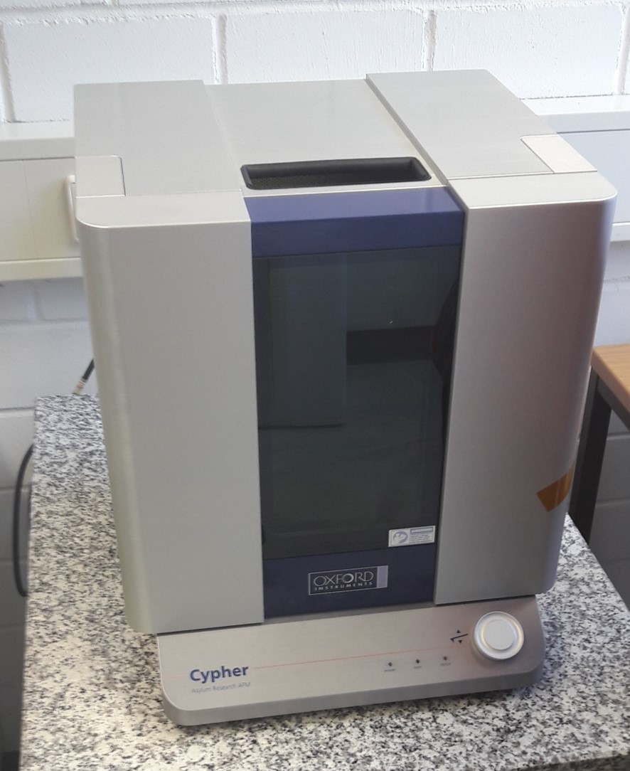
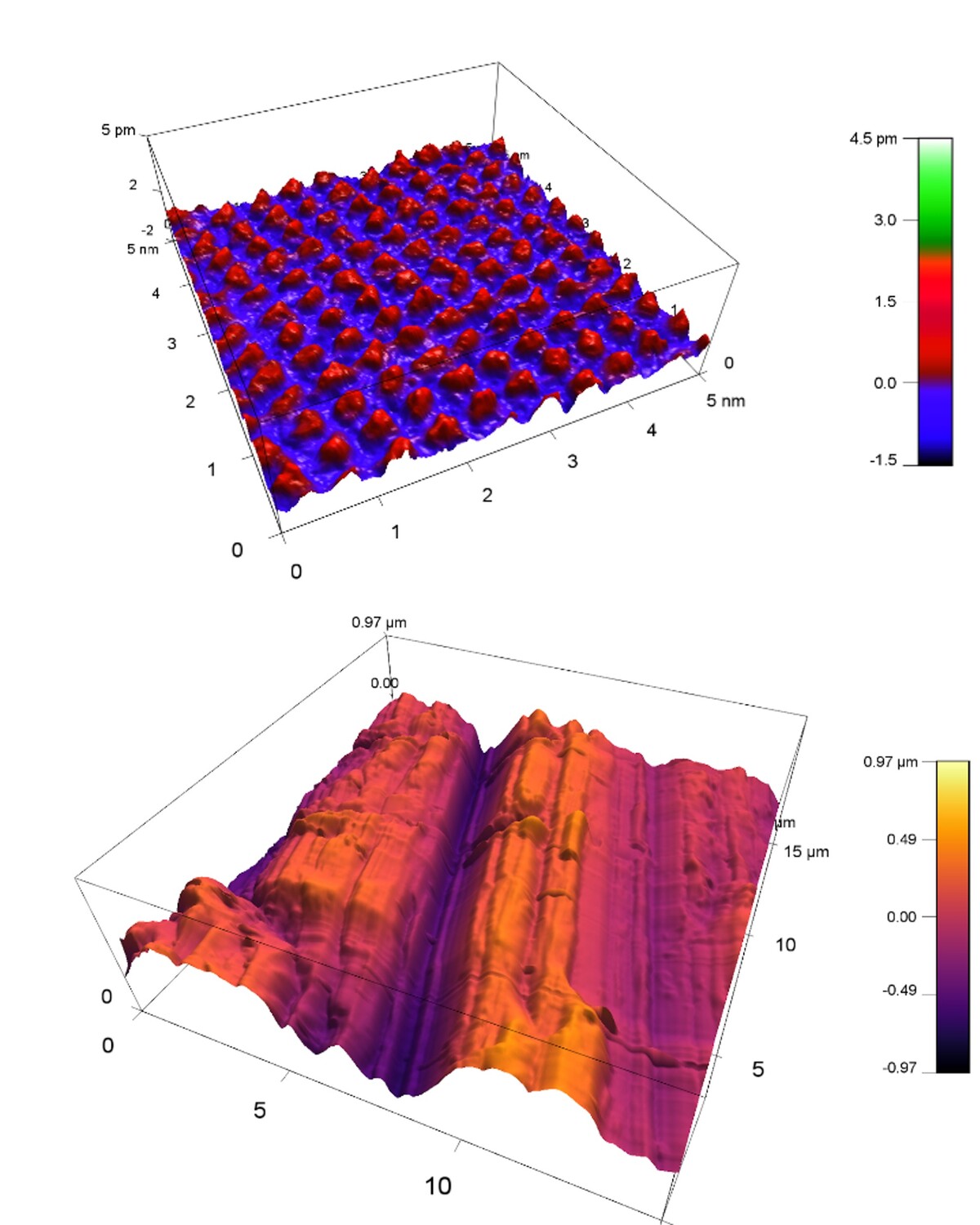
Fig. 1: Growth hillock at the calcite (104) surface observed by contact AFM in situ during crystal growth.

A prerequisite of a detailed understanding of chemical reactions at surfaces are high-resolution microscopic techniques. The atomic force microscope (AFM) gives quantitative insight into surface properties and topography. Advantages of this technique are that no special sample preparation is necessary and measurements with high lateral resolution can be performed at ambient pressure and in solutions.

Surface processes like e.g. crystal growth or dissolution of minerals, can be observed directly in solution in realtime. The incorporation of radionuclides into minerals can be observed and analysed by changes of the crystal growth.

During atomic force microscopy, the surface of the sample is scanned by a nanoscopic sized tip with radius of 2 nm. Spatial resolution in z-direction is about 0.1 nm, and in x,y directions given by the tip radius.

The tip is bonded to a relatively long and elastic bar (ca. 0.1 mm ) with well-known spring constant. The position of the tip is measured by a reflecting laser beam and a segmented photodiode. The bar with the tip is attached to a piezoelectric quartz and moved by electric potentials.

Sensing of the sample surface is performed by essentially two techniques. Either the tip is scanned in permanent contact with the sample surface or the tip oscillates perpendicular to the surface while scanning. If the tip approaches to the surface, attenuation and phase shift of the tip in relation to the exciting piezoelectric quartz occurs, which is detected by the laser and the segmented photodiode. Advantage of the latter technique is that it has less impact onto the sample surface, which is favourable especially in the case of sensitive samples.


Contact:

Dr. Dieter Schild      Dr. Frank Heberling (AFM)     
   +49 721 608 22521 / 22379    +49 721 608 24782